Abstract
Introduction
Multiple myeloma (MM)-induced bone disease remains one of its most devastating complications, caused by increased bone resorption by overactivated osteoclasts coupled with impaired bone formation. MM cells produce osteoclast-activating factors that induce osteoclast activation and extensive bone resorption. Our previous work demonstrated that matrix metalloproteinase 13 (MMP-13) is a critical osteoclastogenic factor that is highly secreted by MM cells (Fu J etc. JCI. 2016). We also identified that the checkpoint inhibitor, programmed death-1 homolog (PD-1H/VISTA), serves as the MMP-13 receptor in osteoclasts and mediates MMP-13-dependent osteoclastogenic function which is largely blocked in Pd-1h -/-osteoclasts (Fu J etc. ASH 2019, 2020). While the inhibitory role of PD-1H/VISTA in T-cells has recently been described (ElTanbouly MA etc. Science 2020), its cellular binding proteins remain unclear, and its role in osteoclast activation andMM bone disease have not been addressed.
Methods and Results
To identify its interacting proteins, PD-1H-His 6 recombinant protein was expressed in mouse bone marrow mononuclear cells, the associated proteins pulled down by Ni-NTA agarose beads from cell lysates and identified by mass spectrometry. Functional annotation charting of the 75 proteins enriched in PD-1H pull-down samples (with signal ratio of PD-1H-His 6 pull-down vs control >2) indicated that almost 30% of the interacting targets were either cytoskeletal or cytoskeleton-associated proteins.
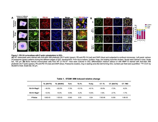
Given that the F-actin cytoskeleton undergoes dynamic reorganization during osteoclast differentiation and plays critical roles in bone resorption, we further addressed the role of PD-1H in F-actin cytoskeleton regulation. Initially, osteoclasts form F-actin-rich adhesive structures, termed podosomes. At later stages, podosomes collectively rearrange into clusters and rings and finally into sealing belts to mediate osteoclast spreading, migration and bone resorption (Teitelbaum SL. Ann N Y Acad Sci. 2011). By confocal immunofluorescence microscopy, we found that PD-1H co-localized with F-actin podosome clusters, rings and sealing belts during osteoclast differentiation (Figure 1A). The functional role of PD-1H in F-actin cytoskeleton reorganization was addressed using Pd-1h -/- osteoclast wherein Pd-1h knockout lead to the disruption of podosome clusters at early stages relative to WT controls, while at later stages, Pd-1h -/-osteoclasts exhibited significantly fewer F-actin rings and belts (Figure 1B). Further, binding of MMP-13 to PD-1H increased the number of osteoclasts forming F-actin rings and belts, as well as the size of F-actin belts, which was blocked in Pd-1h -/- osteoclasts.
To determine the role of PD-1H in the development of myeloma-induced lytic bone lesions, 5TGM1 myeloma cells were bilaterally intratibially injected into Pd-1h wtRag2 -/- or Pd-1h -/-Rag2 -/- mice (n=10) to induce lytic bone lesions. Three weeks following intratibial 5TGM1 injection, tibiae were harvested for micro-computed tomography. Subsequent quantitative histomorphology analyses of the trabecular and cortical bones confirmed that the knockout of Pd-1h reduced MM-induced bone destruction with significantly less decrease in trabecular bone volume (Tb. [BV/TV]), trabecular bone number (Tb. N.), trabecular bone thickness (Tb. Th.), as well as less increase in trabecular bone spacing (Tb. Sp.) and bone specific surface (Tb. [BS/BV]) compared to Pd-1h wtRag2 -/- mice. Similar effects were observed in cortical bone with less decrease in cortical bone thickness (CT. Th.), cortical bone area fraction (CT. [BA/TA]), and cortical tissue mineral density (CT. TMD) in 5TGM1 bearing Pd-1h -/-Rag2 -/- mice vs Pd-1h wtRag2 -/- mice (Table 1).
Conclusions
Taken together, our study, for the first time, reveals the novel role of checkpoint inhibitor, PD-1H/VISTA, in osteoclasts and myeloma bone disease. PD-1H associates with cytoskeleton proteins and regulates the F-actin cytoskeleton reorganization which is critical for osteoclast bone resorption activity. Further, PD-1H mediates MMP-13-induced osteoclast fusion, F-actin belts formation, and osteoclast activation. Pd-1h -/-in recipient mice significantly impairs MM-induced bone loss, demonstrating that PD-1H/VISTA plays a critical role in MM bone disease.
No relevant conflicts of interest to declare.


This feature is available to Subscribers Only
Sign In or Create an Account Close Modal